- 2020 – today
- Partners
- Uniklinik RWTH Aachen (DE)
- Philips Healthcare (NL)
- Futura Composites (NL)
- UMC Utrecht (NL)
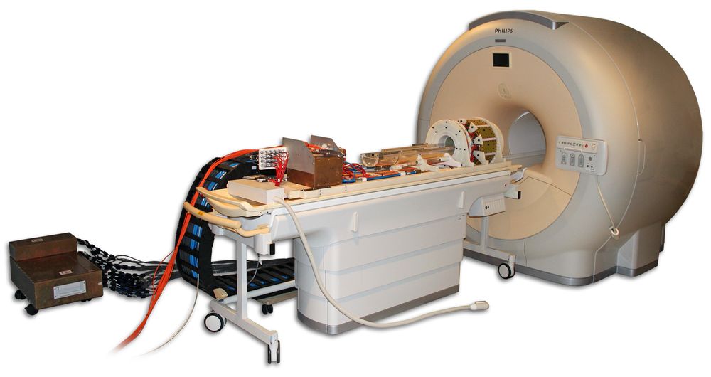
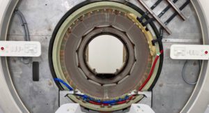
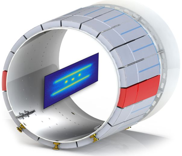
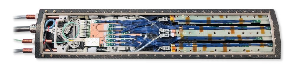
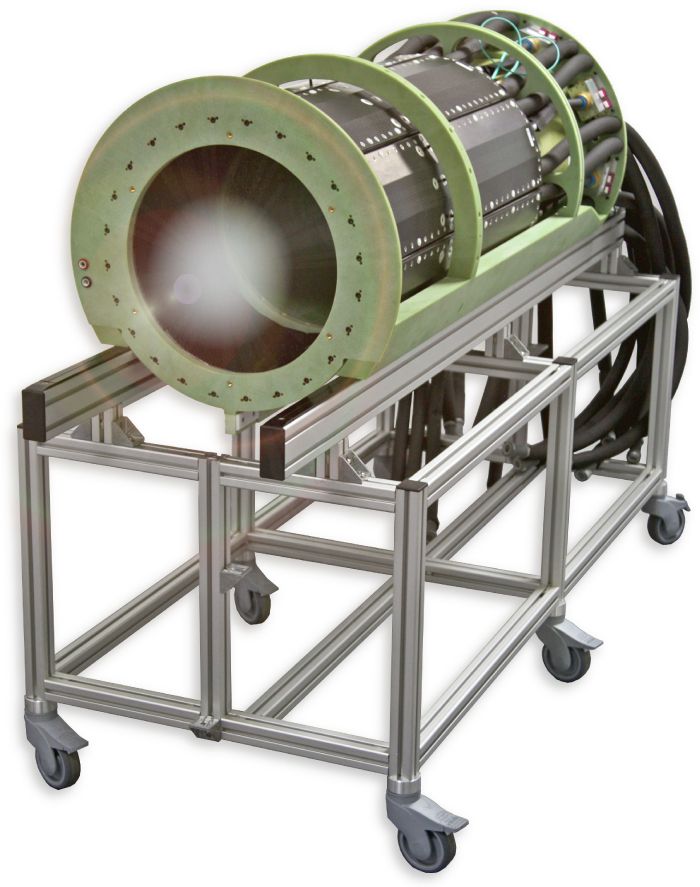
- PET field of view (transaxial × axial): 700 mm × 163 mm (currently 700 mm × 50 mm)
- PET sensor technology: Digital SiPMs DPC 3200-22 (PDPC)
- Total amount of crystals: 15,552 (currently installed: 3,456)
- PET detector: 48×48 mm², single-layer, 12×12 LYSO crystals, 4 mm pitch, 19 mm height
- System: 12 Modules with 9 detector blocks each (currently installed: 8 modules with 3 detector blocks each)
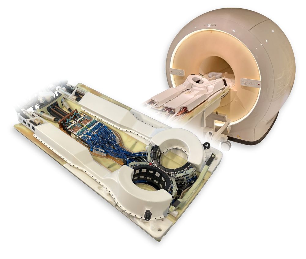
- Target MRI Scanner: Philips Ingenia 1.5T with a split gradient coil
- PET Performance
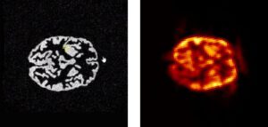
- Energy resolution: 10.9 % FWHM
- Time resolution: 263 ps FWHM
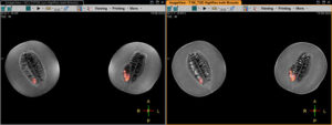
- UMC Utrecht: MRI/PET may better control metastatic tumors
- UMC Utrecht: Part MRI/PET installed. The next step.
- Casper Beijst: Yesterday we had the official handover…
- 2016 – 2022
- Funded by European Union, Horizon 2020 research and innovation programme, grant agreement no. 667211
- Partners
- Uniklinik RWTH Aachen (DE)
- Technische Universiteit Delft / DEMO (NL)
- Futura Composites (NL)
- NORAS MRI Products (DE)
- Philips Electronics (NL)
- Forschungszentrum Jülich (DE)
- Universitätsklinikum Münster (DE)
- Medical University of Vienna (AT)
- European Institute for Biomedical Imaging Research (AT)
- PET field of views:
- Two rings: 2 times 180 mm × 98 mm
- Sternum area is partly covered due to the incliniation of 20° of each ring
- PET sensor technology: digital SiPMs DPC 3200-22 (PDPC)
- Total amount of crystals: 191,800
- Detector: 48×48 mm², 3-layer DOI, 3,425 LSO crystals, 1.33 mm pitch, 15 mm height
- System: Two separate bores with 56 detector blocks arranged on 2 rings
- Target MRI Scanner: Philips Ingenia 1.5T
- MRI RF Coil: 4-Channel Receive Sense Breast Coil
- 2016 – 2021
- A Helmholtz Validation Fund project
- Partners
- Forschungszentrum Jülich GmbH (DE)
- Uniklinik RWTH Aachen (DE)
- Siemens Healthineers (DE)
- Monash University, Victoria, Australia
- Inviscan SA. Strasbourg (FR)
- Affinity Imaging GmbH (DE)
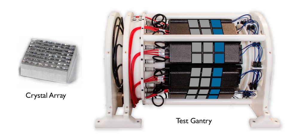
- PET field of view (transaxial × axial): 282 mm × 250 mm
- PET sensor technology: digital SiPMs DPC 3200-22 (PDPC)
- Total amount of crystals: 196,080
- PET detector: 48×48 mm², 3-layer DOI, 1,634 LSO crystals, 2 mm pitch, 24 mm height
- System: 8 Modules with 15 detector blocks each in 5 rings
- Target MRI Scanner: Siemens MAGNETOM Terra 7T
- MRI RF Coil: Custom-made transmit receive head coil
- PET Performance
- Energy resolution: 15 % FWHM (preliminary, to be improved)
- Time resolution: t.b.d ps FWHM
- Sensitivity >12% (simulation)
- Lerche, C., et al. “Development of a UHF-MRI compatible BrainPET insert for neuroscientific applications.” JOURNAL OF CEREBRAL BLOOD FLOW AND METABOLISM. Vol. 42. No. 1_ SUPPL. 2455 TELLER RD, THOUSAND OAKS, CA 91320 USA: SAGE PUBLICATIONS INC, 2022.
- Abstract
DOI:10.1055/s-0040-1708248
Lerche, Christoph, et al. “Design and Simulation of a high-resolution and high-sensitivity BrainPET insert for 7T MRI.” Nuklearmedizin-NuclearMedicine 59.02 (2020): V96.
- 2011 – 2014
- Funding
- of the overall project by the German federal state North Rhine Westphalia (HighTech.NRW)
- of ForSaTum by the European Union, European Regional Development Fund, Investing In Your Future, grant number z0903ht014g
- of Centre of Excellence in Medical Engineering by the Wellcome Trust and EPSRC, grant number WT 088641/Z/09/Z
- Partners
- Uniklinik RWTH Aachen (DE)
- Philips Technologie GmbH (DE)
- King’s College London (UK)
- Ruhr-Universität Bochum
- AplaGen, PharmedArtis
- Kairos
- ITZ Medicom
- Digital Medics
- invivoContrast
- Aachener Kompetenzzentrum Medizintechnik (AKM)
- PET field of view (transaxial × axial): 209.6 mm × 96.6 mm
- PET sensor technology: digital SiPMs DPC 3200-22 (PDPC)
- Total amount of crystals: 54,000
- PET detector: 32×32 mm², single-layer, 30×30 LYSO crystals, 1 mm pitch, 12 mm height
- System: 10 Modules with 6 detector blocks each in three rings
- Target MRI Scanner: Philips Achieva 3T
- MRI RF Coils:
- Large 1H TX/RX coil (rat- to rabbit-size)
- Small 1H TX/RX coil (mouse-size), FOV (transaxial × axial): 46 mm × 120 mm
- Multi-Nuclei 1H/19F coil (rat- to rabbit-size) (transaxial × axial): 100 mm × 100 mm
- Systems built: 3
- PET Performance
- Energy resolution: 12.7 % FWHM [NEMA]
- Time resolution:
- trigger scheme 1: 256 ps [TOF]
- trigger scheme 3: 546 ps [TOF], 605 ps FWHM [NEMA]
- Spatial resolution: 0.9 mm / 0.73 mm³ FWHM [TMI]
- System sensitivity: 4.1 % [NEMA]
- Maximum activity (NECR curve peak): 35 MBq (limited by raw data transmission)
- [TMI]
DOI: 10.1109/TMI.2015.2427993
Weissler, Bjoern, et al. “A digital preclinical PET/MRI insert and initial results.” IEEE Transactions on Medical Imaging 34.11 (2015): 2258-2270.
- DOI: 10.1088/2057-1976/2/1/015010
Düppenbecker, Peter M., et al. “Development of an MRI-compatible digital SiPM detector stack for simultaneous PET/MRI.” Biomedical Physics & Engineering Express 2.1 (2016): 015010.
- DOI: 10.1088/0031-9155/60/6/2231
Wehner, Jakob, et al. “MR-compatibility assessment of the first preclinical PET-MRI insert equipped with digital silicon photomultipliers.” Physics in Medicine & Biology 60.6 (2015): 2231.
- DOI: 10.1016/j.nima.2013.08.077
Wehner, Jakob, et al. “PET/MRI insert using digital SiPMs: investigation of MR-compatibility.” Nuclear Instruments and Methods in Physics Research Section A: Accelerators, Spectrometers, Detectors and Associated Equipment 734 (2014): 116-121.
- DOI: 10.1088/0031-9155/60/6/2231
Wehner, Jakob, et al. “MR-compatibility assessment of the first preclinical PET-MRI insert equipped with digital silicon photomultipliers.” Physics in Medicine & Biology 60.6 (2015): 2231.
- DOI: 10.1088/0031-9155/61/7/2851
Schug, David, et al. “Initial PET performance evaluation of a preclinical insert for PET/MRI with digital SiPM technology.” Physics in Medicine & Biology 61.7 (2016): 2851.
- [NEMA]
DOI: 10.1088/2057-1976/aae6c2
Hallen, Patrick, et al. “PET performance evaluation of the small-animal Hyperion IID PET/MRI insert based on the NEMA NU-4 standard.” Biomedical physics & engineering express 4.6 (2018): 065027.
- [TOF]
DOI:10.1109/TNS.2017.2654920
Schug, David, et al. “Crystal delay and time walk correction methods for coincidence resolving time improvements of a digital-silicon-photomultiplier-based PET/MRI insert.” IEEE Transactions on Radiation and Plasma Medical Sciences 1.2 (2017): 178-190.

- 2010 – 2015
- Funded by European Union FP7, grant agreement no. 241711
- Partners
- Uniklinik RWTH Aachen (DE)
- Philips Research (NL)
- Ruprecht-Karls-Universität Heidelberg (DE)
- TU Delft (NL)
- PET field of view (transaxial × axial): 217.6 mm × 32 mm
- PET sensor technology: digital SiPMs DPC 3200-22 (PDPC)
- Total amount of crystals: 1,280
- PET detector: 32×32 mm², single-layer, 8×8 LYSO crystals, 4 mm pitch, 10 mm height
- System: 10 Modules with 2 detector blocks each in one ring
- Target MRI Scanner: Philips Ingenia 3T
- MRI RF Coil: Large 1H TX/RX coil (rat-to-rabbit size)
- PET Performance
- Energy resolution: 11.4 % FWHM [clinicalSystem]
- Time resolution
- Spatial resolution: 2 mm [clinicalSystem]
- System sensitivity: 0.7 % [clinicalSystem]
- [clinicalSystem]
DOI: 10.1088/0031-9155/60/18/7045
Schug, David, et al. “PET performance and MRI compatibility evaluation of a digital, ToF-capable PET/MRI insert equipped with clinical scintillators.” Physics in Medicine & Biology 60.18 (2015): 7045.
- [TOF]
DOI:10.1109/TNS.2017.2654920
Schug, David, et al. “Crystal delay and time walk correction methods for coincidence resolving time improvements of a digital-silicon-photomultiplier-based PET/MRI insert.” IEEE Transactions on Radiation and Plasma Medical Sciences 1.2 (2017): 178-190.
- 2008 – 2011
- Funded by European Union FP7, grant agreement no. 201651
- Partners
- Philips Technologie GmbH (DE)
- King’s College London (UK)
- Centro Nacional De Investigaciones Cardiovasculares Carlos III (F.S.P.) (ES)
- Stichting Het Nederlands Kanker Instituut-Antoni van Leeuwenhoek Ziekenhuis (NL)
- Universitätsklinikum Hamburg-Eppendorf (DE)
- Ruprecht-Karls-Universität Heidelberg (DE)
- iMinds VZW (BE)
- Fondazione Bruna Kessler (IT)
- PET field of view: 160 mm transaxial x 30 mm axial (94.1 mm prepared)
- PET sensor technology: Analog SiPMs (FBK) with PETA2 ASIC (University of Heidelberg)
- Total amount of crystals: 9,680
- PET Detector: 30×30 mm², single-layer, 22×22 LYSO crystals, 1.3 mm pitch, 10 mm height
- PET System: 10 Modules with 2 detector blocks each in one ring
- Target MRI Scanner: Philips Achieva 3T
- MRI RF Coil: Permanently mounted RF transmit/receive coil (16-rod birdcage resonator)
- FOV (transaxial × axial): 160 mm × 160 mm
- PET Performance
- Energy resolution: 29.7 % FWHM
- Time resolution: 2.5 ns FWHM
- Energy resolution: 29.7 % FWHM
- Spatial resolution: 1.7 mm / 1.8 mm3 FWHM
- System sensitivity: 0.6 %
- Maximum activity: 35 MBq

- DOI:10.1088/0031-9155/59/17/5119
Weissler, Bjoern, et al. “MR compatibility aspects of a silicon photomultiplier-based PET/RF insert with integrated digitisation.” Physics in Medicine & Biology 59.17 (2014): 5119.
- DOI:10.1109/TNS.2015.2392560
Mackewn, Jane E., et al. “PET performance evaluation of a pre-clinical SiPM-based MR-compatible PET scanner.” IEEE Transactions on nuclear science 62.3 (2015): 784-790.
- DOI:10.1109/NSSMIC.2013.6829111
Soultanidis, Georgios M., et al. “Demonstration of motion correction for PET-MR with PVA cryogel phantoms.” 2013 IEEE Nuclear Science Symposium and Medical Imaging Conference (2013 NSS/MIC). IEEE, 2013